Hoffman EA, Reinhardt JM, Sonka M et al (2003) Characterization of the interstitial lung diseases via density-based and texture-based analysis of computed tomography images of lung structure and function. Kim HG, Tashkin DP, Clements PJ et al (2010) A computer-aided diagnosis system for quantitative scoring of extent of lung fibrosis in scleroderma patients. doi: 10.1097/RCT.0b013e31818da65cĬamiciottoli G, Orlandi I, Bartolucci M et al (2007) Lung CT densitometry in systemic sclerosis: correlation with lung function, exercise testing, and quality of life. Sumikawa H, Johkoh T, Yamamoto S et al (2009) Computed tomography values calculation and volume histogram analysis for various computed tomographic patterns of diffuse lung diseases. Lynch DA (2007) Quantitative CT of fibrotic interstitial lung disease. doi: 10.1164/rccm.200802-304EDĬollins CD, Wells AU, Hansell DM et al (1994) Observer variation in pattern type and extent of disease in fibrosing alveolitis on thin section computed tomography and chest radiography. Strange C, Seibold JR (2008) Scleroderma lung disease: “if you don’t know where you are going, any road will take you there”. LeRoy EC, Black CM, Fleischmajer R et al (1988) Scleroderma (systemic sclerosis): classification, subsets and pathogenesis.
#Osirix dicom free software#
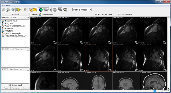
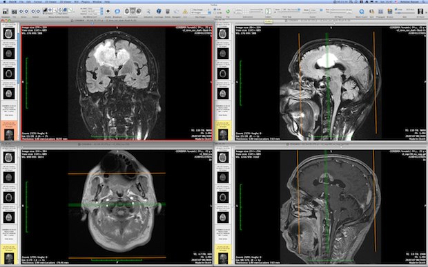
Rosset A, Spadola L, Ratib O (2004) OsiriX: an open-source software for navigating in multidimensional DICOM images. J Comput Assist Tomogr 30:244–249īest AC, Lynch AM, Bozic CM et al (2003) Quantitative CT indexes in idiopathic pulmonary fibrosis: relationship with physiologic impairment. Sumikawa H, Johkoh T, Yamamoto S et al (2006) Quantitative analysis for computed tomography findings of various diffuse lung diseases using volume histogram analysis.

Sverzellati N, Zompatori M, De Luca G et al (2005) Evaluation of quantitative CT indexes in idiopathic interstitial pneumonitis using a low-dose technique. Zavaletta VA, Bartholmai BJ, Robb RA (2007) High resolution multidetector CT-aided tissue analysis and quantification of lung fibrosis. J Rheumatol 18:1520–1528Ĭozzi F, Chiesura Corona M, Rizzi M et al (2001) Lung fibrosis quantified by HRCT in scleroderma patients with different disease forms and ANA specificities. Warrick JH, Bhalla M, Schabel SI, Silver RM (1991) High resolution computed tomography in early scleroderma lung disease. Goldin JG, Lynch DA, Strollo DC et al (2008) High-resolution CT scan findings in patients with symptomatic scleroderma-related interstitial lung disease. Wells AU (2008) High-resolution computed tomography and scleroderma lung disease. Goh NSL, Desai SR, Veeraraghavan S et al (2008) Interstitial lung disease in systemic sclerosis: a simple staging system. Karassa FB, Ioannidis JPA (2008) Mortality in systemic sclerosis. Steen VD, Conte C, Owens GR, Medsger TA (1994) Severe restrictive lung disease in systemic sclerosis. The study provides the new working hypothesis that OsiriX may be a useful and feasible tool to achieve a quantitative evaluation of PF in SSc patients. A significant difference between the mean time spent on the OsiriX quantitative analysis (mean 1.85 ± SD 1.3 min) and the mean time spent by the radiologist for the HRCT semiquantitative assessment (mean 8.5 ± SD 4.5 min, p < 0.00001) was noted. An excellent intra-reader reliability of HRCT findings among both readers was obtained. Moreover, kurtosis correlated significantly with the mean lung attenuation (Spearman’s rho = 0.885 p = 0.0001). A significant association between the median values of kurtosis by both the quantitative OsiriX assessment and the HRCT semiquantitative analysis was found ( p < 0.0001). Intra-reader reliability of HRCT findings and feasibility of OsiriX quantitative segmentation was recorded. The results obtained were compared with those of HRCT semiquantitative analysis. parameters of distribution of lung attenuation such as kurtosis and mean lung attenuation) of PF was independently performed on the same sections by a rheumatologist, independently and blinded to radiologists’ scoring, using OsiriX. For the assessment of the extension of PF, the adopted semiquantitative HRCT score ranged from 0 to 3 (0 = absence of PF 1 = 1–20 % of lung section involvement 2 = 21–40 % of lung section involvement 3 = 41–100 % of lung section involvement).

Pulmonary involvement was evaluated in three sections (superior, middle and inferior). Chest high-resolution computed tomography (HRCT) examinations obtained from 10 patients with diagnosis of SSc were analysed by two radiologists adopting a standard semiquantitative scoring for PF. To investigate the utility of an open-source Digital Imaging and Communication in Medicine viewer software-OsiriX-to assess pulmonary fibrosis (PF) in patients with systemic sclerosis (SSc).


 0 kommentar(er)
